3.1 Open the file 'insulin.pdb' or click here.
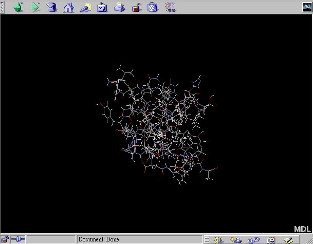
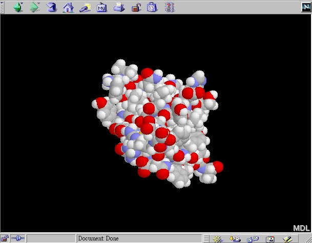
3.2 Try to change the display mode to 'Spacefill'. Do you remember how to do this?
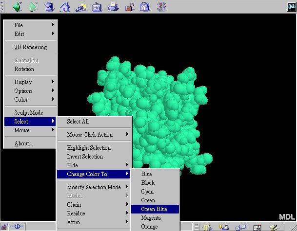
3.3 Instead of using the default color setting, you can color the molecule differently. Try the following to change the color to green-blue:
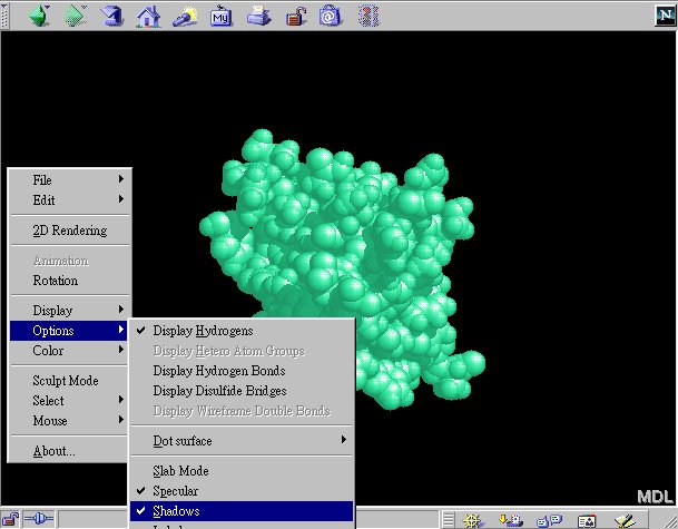
3.4 You can make the view better looking by casting a shadow and selecting the 'specular' mode.
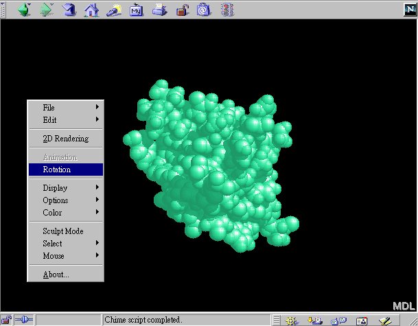
3.5 You can turn on/off the rotation of the molecule by:
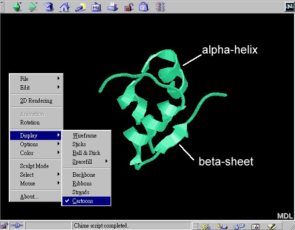
3.6 Another useful representation of protein is the 'Cartoons' mode. In the representation, alpha helices are shown as ribbons while beta sheet are shown as arrows:
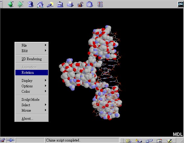
3.7 Open the file 'gal4.pdb' or click here. The complex consists of protein and DNA. Then how do you know which part is DNA and which part is protein?
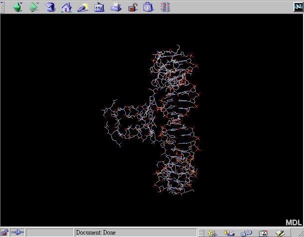
3.8 You can first select the protein by:
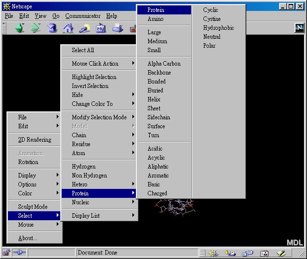
3.9 Then change the display mode of protein to 'spacefill'. Notice only the protein part of the complex is shown in 'spacefill' mode and the DNA part is shown in 'wireframe mode'. You can see it more clearly by turning on 'rotation'.