
Teacher Development Course
Using molecular modeling software to view protein structures
Presented by Kam Bo Wong
Department of Biochemistry & Molecular Biotechnology Programme
email: kbwong@cuhk.edu.hk
http://smart.bch.cuhk.edu.hk/kbwong/index.htm
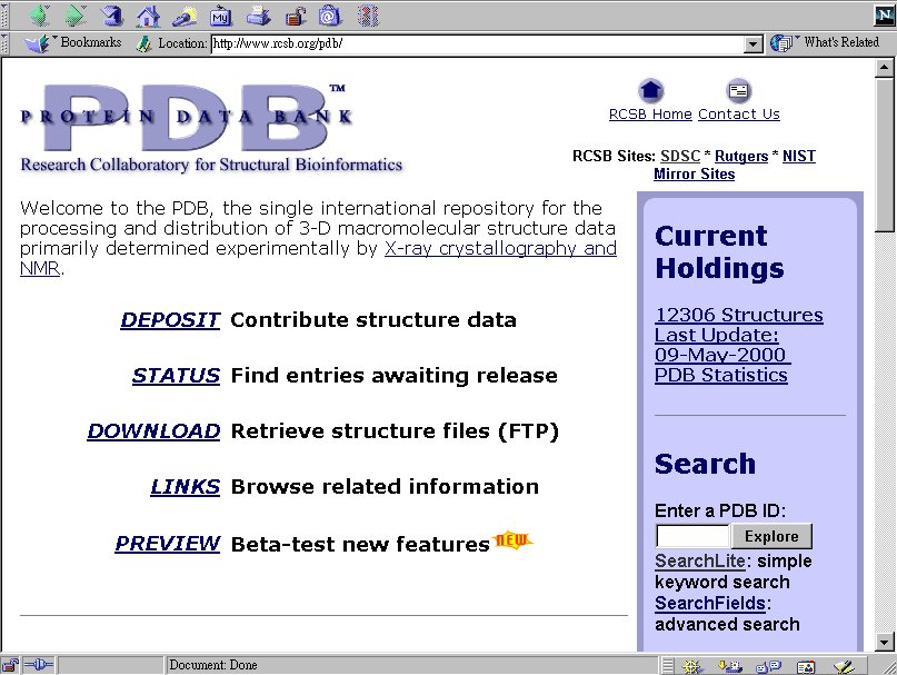
4. Where to obtain protein structure files?
Protein structure files can be downloaded from the Protein
Data Bank. All entries in the PDB is identified by a four-letter code.
The PDB code for the insulin is '3ins'. If you don't know the PDB code
for a protein, you can click the 'SearchLite' to search for it.
4.1 Go to the PDB home page: http://www.rcsb.org/pdb/
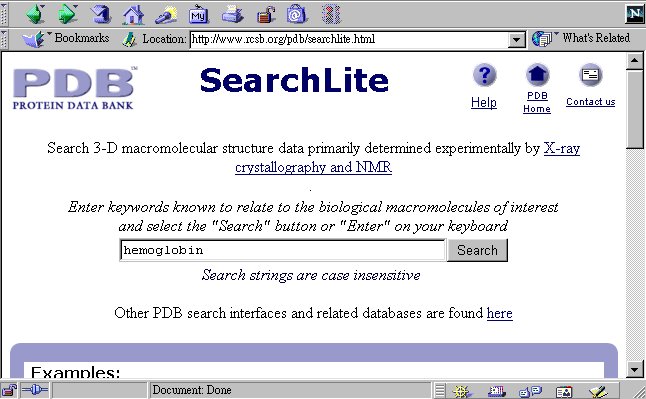
4.2 Click SearchLite to search for the structure file you want.
Try type hemoglobin and press the search button.
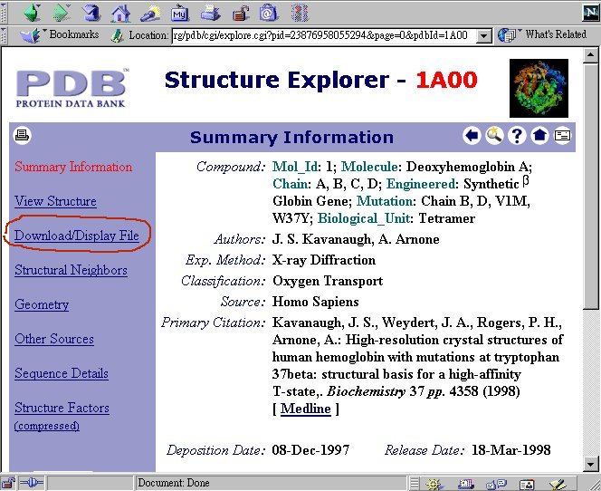
4.3 You will see a number to hits. They are structures of hemoglobin
and its variants (hemoglobin is one of the most well studied proteins).
Notice all structures in the Protein Data Bank are identified by a four-letter
code (e.g. 1A00). Now click 'EXPLORE' on the left of the '1A00' entry.

4.4 You are presented with a summary information of the protein
you have just selected. Click the 'Download/Display File' on the left column:
4.5 Select the format of the structure file and save the structure
in your local hard disk.
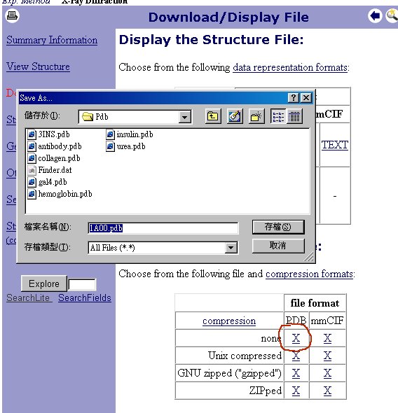 4.6 Now you have download a pdb file for hemoglobin. Try to practice
what you have learnt today and use Chime to view the 3D structure of this
file. (If the internet connection is too slow, there is a local copy of
the file 1A00.pdb here.
4.6 Now you have download a pdb file for hemoglobin. Try to practice
what you have learnt today and use Chime to view the 3D structure of this
file. (If the internet connection is too slow, there is a local copy of
the file 1A00.pdb here.