Viewing Small Organic Molecule
(Download urea.pdb)
The following skill will be taught here:
¡@
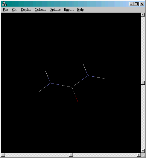
Once you open up a ".pdf" file, e.g. a urea molecule, it will look like this:

¡@(Download human amylase.pdb)
You can change the appearance of the molecule by clicking the 'Display' menu from the toolbar. The default display mode is 'wireframe'. You can also choose the following display:
|
Display |
¡@ |
| Wireframe(default) | Only bond between atoms are shown |
| Backbone |
Representation of a polypeptide backbone as a series of bonds connecting the adjacent carbons of each amino acid in a chain |
| Sticks | Atoms are represented by thick lines |
| Spacefill | Atoms are represented by spheres with size dependent on their atom type |
| Ball & Stick | Atoms are represented by spheres and bonds are represented by thicker lines |
| Ribbons | Structures are represented by a smooth solid "ribbon" surface passing along the backbone |
| Strands | Structures are represented by parallel strands of depth-cued curves passing along the backbone |
| Cartoons | Display of the cartoon version of the 'ribbons' display |
(Examples can be seen by clicking the name of the display in the above table.)
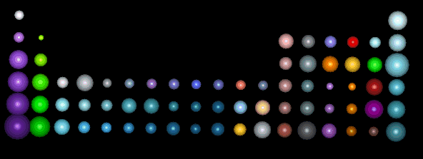
There is an assigned colour for each atom. In the default colour setting, hydrogen atoms are white, nitrogen atoms are blue and oxygen atoms are red. Other colours of the atoms can be found in the following periodic table:

By using the mouse, you can do the following:
Rotate the molecule - Press the left mouse button and dragging the mouse
Move the molecule - Press the right mouse button and dragging the mouse
Zoom in/ out - Press the SHIFT key AND the left mouse button simultaneously. The molecule will be bigger if the mouse moves upwards and be smaller if the mouse moves downwards