Research Highlight

1. Stem Cell Biology and Cardiovascular Regeneration (幹細胞生物學與心血管再生)


Discovery of novel determinants of endothelial lineage using chimeric heterokaryons

Endothelial cells form the inner surface of blood vessels, acting like a non-stick coating. In addition to making substances that keep blood from sticking to the vessel wall, endothelial cells generate compounds that relax the vessel, and prevent it from thickening. Endothelial cells also form capillaries, the smallest vessels that provide oxygen and nutrients for all tissues. A regenerating organ, or a bioengineered tissue, requires a system of capillaries and other microvessels. Thus, regenerative medicine could benefit from a knowledge of how to generate endothelial cells from pluripotent stem cells – cells that can “differentiate” to form almost any type of cell in the body.

We have used a cell fusion model (named heterokaryon) to track the changes in gene expression that occur as a pluripotent stem cell differentiates to ultimately become an endothelial cell. In this model, mouse embryonic stem cells (ESCs) are fused to human endothelial cells. Over time the human endothelial cells drive gene expression in the ESCs toward that of endothelial cells. We recently discovered changes in gene expression in many genes that have not previously been described as involved in the differentiation of endothelial cells. When one of these genes – named Pou3f2 – was inactivated in ESCs, they could not be differentiated into endothelial cells. The absence of Pou3f2 also drastically impaired how blood vessels developed in zebrafish embryos. Thus the heterokaryon model can generate important information regarding the dynamic changes in gene expression that occur as a pluripotent cell differentiates to become an endothelial cell. This model may also be useful for discovering other genes that control the differentiation of other cell types. This novel finding has just been published in Elife (2017 Mar 21;6. pii: e23588. doi: 10.7554/eLife.23588.) [Pudmed] https://elifesciences.org/articles/23588

Representative Figure 1. Heterokaryon recapitulates gene expression of endothelial ontogeny.
Mobirise


2. Nanotechnology and Vascular Biology (納米科技與心血管生物學)


E-selectin-targeting delivery of microRNAs by microparticles ameliorates endothelial inflammation and atherosclerosis

Nanoparticles have emerged as a promising vector for miR delivery, and polyethylene glycol-polyethyleneimine (PEG/PEI) nanoparticles have been validated as an efficient delivery vector. However, rapid clearance and poor targeting limit their clinical value. We developed and tested a porous silicon multistage vector (MSV) delivery system, which is a micrometer-sized nanoporous microparticle. Large amounts of therapeutic agents have been packaged into PEG/PEI nanoparticles and loaded into the nanopores within the MSV microparticles.

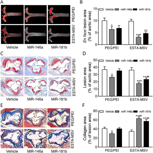
E-selectin is a surface marker of endothelial cell (EC) inflammation, one of the hallmarks of atherogenesis. Thus, we tested the hypothesis that delivery of microRNA (miR)-146a and miR-181b with an E-selectin-targeting multistage vector (ESTA-MSV) to inflamed endothelium covering atherosclerotic plaques inhibits atherosclerosis. Cy5-conjugated miR-146a and miR-181b were packaged in polyethylene glycol-polyethyleneimine (PEG/PEI) nanoparticles and loaded into ESTA-MSV microparticles. Both miRs were downregulated in tumor necrosis factor (TNF)-α-treated ECs. Transfection of TNF-α-treated mouse aortas and cultured ECs with miRs was more efficient with ESTA-MSV than with the PEG/PEI. Likewise, miR-146a/-181b packaged in ESTA-MSV efficiently suppressed the chemokines, CCL2, CCL5, CCL8, and CXCL9, and monocyte adhesion to ECs. Complementary in vivo tests were conducted in male apolipoprotein E-deficient mice fed a Western diet and injected intravenously with the particles prepared as above biweekly for 12 weeks. Treatment with miRs packaged in ESTA-MSV but not in PEG/PEI reduced atherosclerotic plaque size. Concurrently, vascular inflammation markers, including macrophages in aortic root lesions and chemokine expression in aortic tissues were reduced while the vascular smooth muscle cells and collagen increased in plaques from ESTA-MSV/miRs-treated vs. vehicle-treated mice. Our data supported our hypothesis that ESTA-MSV microparticle-mediated delivery of miR-146a/-181b ameliorates endothelial inflammation and atherosclerosis. This novel finding has just been published in Sci Rep (2016; 6: 22910.). [Pudmed] https://www.ncbi.nlm.nih.gov/pmc/articles/PMC4783714/

Representative Figure 4: Effects of PEG/PEI/miRs and ESTA-MSV/miRs on the aortic atherosclerosis of ApoE−/− mice.
Mobirise

3. Metabolic Syndrome and Nutritional Sciences (代謝綜合徵和營養科學)


Adiponectin is required for PPARγ-mediated improvement of endothelial function in diabetic mice.

Obesity and diabetes are common risk factors for the initiation of vascular dysfunction. Adipose tissue is now recognized as an important metabolic and endocrine organ in the regulation of glucose metabolism. Dysregulation of adipose tissue participates in the development of insulin resistance and vascular complications of diabetes.
Rosiglitazone is a PPARγ agonist commonly used to treat diabetes. In addition to improving insulin sensitivity, rosiglitazone restores normal vascular function by a mechanism that remains poorly understood. Here we show that adiponectin is required to mediate the PPARγ effect on vascular endothelium of diabetic mice. In db/db and diet-induced obese mice, PPARγ activation by rosiglitazone restores endothelium-dependent relaxation of aortae, whereas diabetic mice lacking adiponectin or treated with an anti-adiponectin antibody do not respond. Rosiglitazone stimulates adiponectin release from fat explants, and subcutaneous fat transplantation from rosiglitazone-treated mice recapitulates vasodilatation in untreated db/db recipients. Mechanistically, adiponectin activates AMPK/eNOS and cAMP/PKA signaling pathways in aortae, which increase NO bioavailability and reduce oxidative stress. Taken together, these results demonstrate that adipocyte-derived adiponectin is required for PPARγ-mediated improvement of endothelial function in diabetes. Thus, the adipose tissue represents a promising target for treating diabetic vasculopathy. This novel finding has just been published in Cell Metabolism (2016; 6: 22910.).
[Pudmed] https://www.ncbi.nlm.nih.gov/pubmed/21723508

Representative Figure 1. Adipose Tissue Is Required for PPARγ Activation-Induced Amelioration of the Impaired Endothelium-Dependent Relaxations in db/db Mouse Aortae.
Mobirise

Powered by Modeve Limited